


Scientific journal
Fundamental research
ISSN 1812-7339
"Перечень" ВАК
ИФ РИНЦ = 1,798
INNOVATIVE METHODS OF NANOMATERIAL GRAFTS APPLICATION IN NEUROSURGERY

Many nanomaterials possess unique properties. Let us consider properties of an any material particle sized kind of tens and hundreds nanometers. In such a nanoparticle compared to larger objects the percentage of surface atoms or molecules increases in comparison with total atoms (molecules). It influence the given particle properties as a whole. Electric, magnetic, mechanic and some other properties of the material composed of such nanoparticles are no longer constant and begin depending on the size and form of the particles. Sometimes the nanostructured materials show absolutely surprising qualities, which can find potential application in the most various branches of science and technology, in medicine particularly.
In the Belgorod State University an integral method of getting biocomposites based on nanostructured titanium and its alloys with nanocrystalline bioactive calcium-phosphate coatings for making medical grafts. The presence of the coating gives the composite bioactive properties, that improves the graft ability to integrate with bone marrow cells and form bony tissue at the coatings surface. The coatings of nanocrystalline bone-salt, for which an ingenious method of synthesis and application to the graft surface has been developed, is of special interest.
In connection with this, the purpose of our research has been the study of biocompatibility and regenerative processes of bony tissue at operational interventions into bony tissue using materials with nanocrystalline bone-salt coatings.
Materials and methods
Model implants of nanostructured technically pure titanium of the trademark BT1-0 were produced in the form of a rod. Using the micro-arch oxidizing method the implant was covered with a layer of 2-5mcm thick and controlled porosity titanium dioxide.
Then nanocrystalline high-purity bone-salt [1] with layer thickness of about 5mcm was applied to the surface.
Laboratory animals (20 "V-star"-line rats) were involved into the experiment. The animals were divided into the following groups. 15 rats were subjected to resection craniotomy in the right temporal region with the implantation of a nanostructured titanium graft coated with nanocrystalline bony-salt. 5 animals made the control group (false-operated).
The rats were given ether narcosis. After the treatment of the operative field in aseptic conditions a 0,8cm length incision of soft tissues up to the bone was made. The wound edges were separated. In the region of the transitory fold a widowing was performed with an ophthalmic scalpel. The opening was enlarged up to the graft size, i.e. to 0,3x0,5cm. The implants were imbedded into the grade without biostructures´ entrapment. The wound was sewed tightly. The toilette was carried out by means of brilliant green solution. After the anaesthesia recovery the rat is active, performs the toilette of its external integuments. Active movement retained in full, there were no pyramidal insufficiency signs registered.
After the operation the rats were divided into 3 groups: the regeneration study in 7 days (the 1st group), the regeneration study in 14 days (2), the regeneration study in 21 days (3), 45 days (4).
On the preset terms the animals were beaten death by decapitation against the background of etherization. For the determination of possible graft toxicity the parenchymal organs (liver, kidneys, lungs, heart) were taken out for the macro-examination and micro-histological study.
Black and grey rabbits weighing 2-2,5 kg (6)were used. 150 ml of thiopental were administered intramuscularly as narcosis. After sleeping phase coming-in an up to 4 cm length incision of soft tissues was performed in the right frontotemporal region in aseptic conditions. The bone was skeletonized and windowed, the opening being formed into a square burr hole sized 1,0x1,0 cm. The defect was substituted by a metal graft with nanocoating. The graft was fixed in the defect by jointing on the muscle and periosteal coverage. The skin was sutured. The animals were killed in 7 and 14 days.
The bone lamella was taken out together with the graft, examined and photographed through the scanning electron microscope FEI Quana 200 3D without additional treatment. The histological materials were colored with hematoxylin and eosin and subjected to a research under the optical microscope "TOPIS-T" CETI.
The bone lamella was taken out together with the graft, examined and photographed through the scanning electron microscope FEI Quana 200 3D without additional treatment.
Research results
At the function study of the animals it is evident that cognitive and mainly neurophysic functions are not altered. The animals are active, vigorous in the open plain, active movements are retained in full. There are no changes registered on the part of the cardiovascular, respiratory and digestive systems.
At the microscopic examination of the animals it is seen that the incisions were healed by means of primary intension. By the 21st day the traumatic defect didn´t stand out from the general surface. It should be noted that in the animals, having been operated using experimental implants, the vascular pattern is more distinct than that of the false-operated ones. Besides, in the experimental animals the dura mater vessels´ frank repletion in the region adjacent to the bone lamella was defined on the 7th and 14th days.
At the submicroscopic scanning the following processes were described by us. So, in 7 days we observed the filling of the defect between the retained bony tissue and the implanted graft with argyrophil and collage fibers, which were already located on the lamella itself, they filling empty spaces with their processes in the regions with calcium-phosphate coating (Fig. 1). When studying the animals in 14 days, the covering of the whole lamella from the outside with a heavy layer of collagen and elastinic fibers occurs. The fibroblasts with deviating from them collagen fibers are seen very well. Only insignificant regions with the calcium-phosphate coating implant baring or fragments covered with argyrophil fibers, as analogous to the animals killed at the 7-day regeneration stage are defined.
When studying the reparation processes in 21 days, a panniculus, which is a rough fibrous tissue represented by collagen fibers located loosely and disorderly with the intercellular matrix, is seen, next is a lamellar bony tissue, wherein collagen fibers are located in parallel rows (bone lamella), but the orientation of the fibers in the neighboring layers is different. The lamellar bony tissue forms compact and spongy bone layers. The compact layer defines the mechanical strength of the bone and consists of lamellar bone tissue, where blood vessels and nerves begin being formed, and osteons. The spongy layer, which is inside the bone, only starts being formed. The lamellar bone tissue fibroblasts with greater amount of collagen and elastinic fibers gradually oust the cells of the osseous system process layer: osteoblasts, osteocytes and osteoclasts.
There is a great amount of processes, with the help of which they make contacts with neighboring cells, in the osteoblasts. Secreted by practically all the cell´s surface, procollagen contacts actively with the nanocoating. It should be noted that of the two types of osteoblasts (active and inactive) their active forms, which are responsible for the synthesis of collagen and other proteins being part of the organic bone matrix, deposit and exchange of calcium and other ions, occur for the most part.
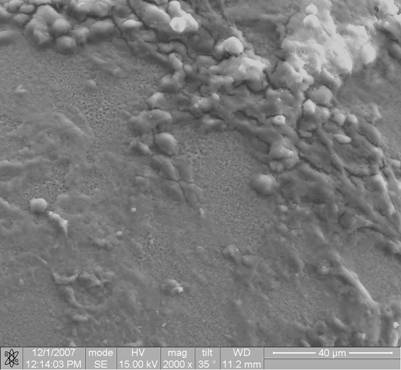
Fig. 1.А
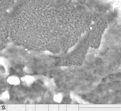
Fig. 1.B
Fig. 1. Titanium alloy graft fragment with nano-bone-salt coating when implanted into the rat´s scull bony tissue (45 days of exposition). Covering of the graft with cells.
SEM. Fig. 1.A. PM (power magnification) x 2000. Fig. 1.B. Fragment of Fig. 1.A. PM x 5000.
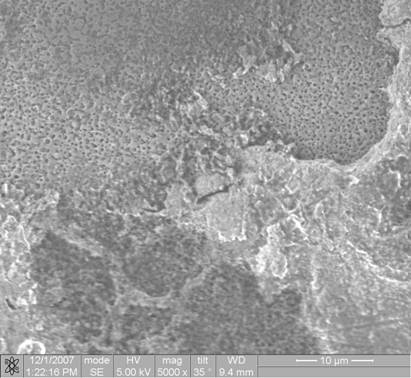
Fig. 2.A
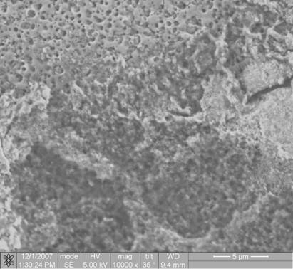
Fig. 2.B
Fig. 2. Titanium alloy graft fragment with nano-bone-salt coating when implanted into the rat´s scull bony tissue (45 days of exposition). Cells´ processes growth into the graft coating.
SEM. Fig. 2.A. PM x 5000. Fig. 2.B. Fragment of Fig. 2.A. PM x 10000.
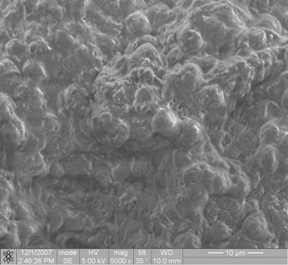
Fig. 3.A
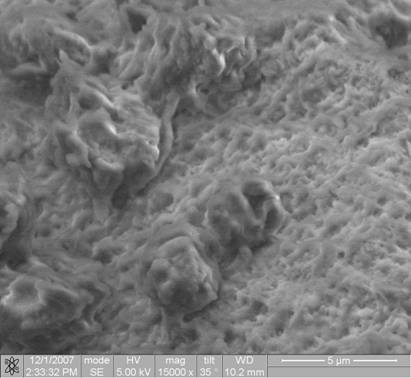
Fig. 3.B.
Fig. 3. Titanium alloy graft fragment with nano-bone-salt coating when implanted into the rat´s scull bony tissue (45 days of exposition). Filling of the graft with fibrous tissue and cells.
SEM. Fig. 3.A. PM x 5000. Fig. 3.B. Fragment of Fig. 3.A. PM x 15000.
Very few osteocytes occurred at this stage. The tissue was not yet fully structured and deprived of lacunes. The osteocytes represented round shape cells with long fine processes. The osteocytes in the basal region contained nuclei, many mitochondria, granular endoplasmic reticulum elements, Golgi complex. Moreover, a lamellar bone tissue formed by bone lamellas was defined in the preparations, it forming compact and spongy substance in the bone.
It should be remarked that between the first and 3-4 days the formation of a haematoma in the graft place was gross observed. Then, by the 7-14th day together with mildly expressed inflammation we observed the migration and proliferation of mesenchymal cells, the formation of fibrovascular tissue round the implant. After that the vascular invasion into the graft, osteoplastic resorption of the last and formation of a neoformed bone on the implant´s surface took place.
At the examination of the rats in 45 days it was shown that the most part of the lamella was substituted by the tissue analogous to bone one located near the region of trepanation. When compared, the regeneration in rabbits occurred more quickly than that in the rats.
At the study of the parenchymal organs (liver, kidneys, lungs, heart) in a week after the implant introduction mildly expressed repletion was defined, that is indicative at the given stage of the false-operated animal group as well. There were no changes in 14 and 21 days registered.
Thus, the operative treatment using titanium implants with calcium-phosphate nanocrystalline bony-salt coatings helps better regeneration of bone tissue, the intoxication phenomena and nanopathology development being not found out. The use of innovation methods of allotransplantology makes quick and noninvasive repair of bony structures possible.
References:
- Verzen R. Preparation of demineralized bone matrix for clinical use // Demineralized bone graft and its application. SPb., 1993 - pp. 4-11.
- Patent application RF № 2007130861 "Method of nanosized bone-salt obtaining", Volkovnyak N.N., Ivanov M.B., Kolobov Yu.R., Buzov A.A., Chuyev V.P.
- Kovalevsky A.M., Iordanishvili A.K. Abolition parodontal and periapical infection combined loci // Urgent problems of clinics, diagnostics and therapy. - SPb., 1995 - pp. 114-115.
- Leontyev V.K. Biologically active synthetic calcium-phosphate-containing materials for oral medicine // Stomatology - 1996 - N5 - pp. 4-6.
- Pavlova T.V., Pavlova L.A., Pavlov I.A. Response of living cells tissues to titanium-aluminium-vanadium alloys implantation // System analysis and management in biomedical systems. - 2007 - N2, Vol.6 -
pp. 364-365.
Библиографическая ссылка
T.V. Pavlova, V.V. Krivetsky, L.A. Pavlova, M.B. Ivanov, D.A. Kolesnikov, Yu.R. Kolobov, I.A. Pavlov INNOVATIVE METHODS OF NANOMATERIAL GRAFTS APPLICATION IN NEUROSURGERY // Фундаментальные исследования. 2009. № 3. С. 40-45;URL: https://fundamental-research.ru/en/article/view?id=1878 (дата обращения: 02.05.2026).